The small pieces of corneal stroma removed during refractive surgery can serve as a scaffold for growing corneal endothelium.
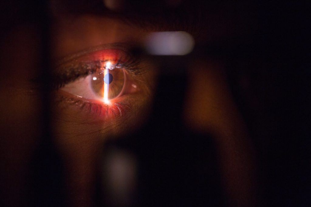
Defects in the corneal endothelium can be assessed in part using a slit-lamp examination.
The cornea is one of the most commonly transplanted tissues worldwide, and it also has one of the lowest rejection rates. This is due to the fact that the cornea is avascular. Donor corneas are also easier to harvest compared to other tissues. Nevertheless, the donor corneas are in short supply. The most common indication for a corneal transplant is to replace the corneal endothelium that has been dysfunctional in pumping out excess water, leading to cloudy corneas.
This is why current research tries to find ways to engineer the endothelial layer to provide an alternative to donor corneas. In the past, there have been several synthetic and biological materials used to construct the corneal endothelium. One of the biologic materials speculated for us are the lenticules extracted during retractive surgery, which have corneal stroma and are more readily available than donor corneas. These lenticules are promising because they have been used as implants successfully to treat errors of refraction, tears, and perforations.
This study was defined as a proof-of-concept study to demonstrate the suitability of using decellularized lenticules for culturing and transplanting the corneal endothelium. Decellularization is necessary to remove remaining cellular material that can trigger an immune response. However, at the same time, it is important that the native extracellular matrix is preserved as much as possible, as this will be the scaffold that regulates cellular differentiation and tissue remodeling in the host. The corneal endothelium matrix should also be optically clear.
In this experiment, lenticules from SMILE (small-incision lenticule extraction) surgery were obtained and decellularized by incubation in a culture medium for corneal endothelium. Decellularization was also confirmed by evaluating DNA content, gel electrophoresis, and microscopy with DAPI staining. Lenticule transparency was ascertained by calculating the modulation transfer function, and the content of the extracellular matrix was determined by checking for collagens and glycosaminoglycans. Afterwards, corneal endothelium cells obtained from donor corneas that were deemed not good for transplant were cultured and allowed to grow on the decellularized lenticules. The engineered tissues were then tested for functionality through an ex-vivo transplant model, in which they were transplanted into edematous donor corneas and perfused artificially for 10 days.
Results showed that after decellularization, the amount of DNA per dry tissue weight of the lenticules was significantly reduced. In addition, the method used for decellularization also did not change the transparency or the arrangement of the collagen fibers in the extracellular matrix. In addition, the corneal endothelium cells attached to this scaffold and eventually matured to express significant proteins associated with differentiated cells. The engineered tissue was also found to significantly reduce corneal edema in the ex-vivo transplant model, and it was noted that the corneal endothelial cells migrated to the recipient cornea that was without and repopulated the latter.
There are, however, disadvantages to using SMILE lenticules. The first is that they tend to be small, only around 6 millimeters on average, and so will not be useful if larger tissues are necessary. This is also implies that that there is little allowance for dislocation or decentration of the lenticule post transplant. It is also possible that, because of the tissue thickness, transplanting this may case a hyperopic shift (farsightedness).
Nevertheless, all of these findings demonstrate that this techniques of using decellularized SMILE lenticules, accomplished with a chemical-free-method, is feasible and suitable for constructing the corneal endothelium for transplantation.
Hazra, S. et al. (2022) “Use of decellularized smile (small-incision lenticule extraction) lenticules for engineering the corneal endothelial layer: A proof-of-concept,” Current Eye Research, 48(3), pp. 251–262. Available at: https://doi.org/10.1080/02713683.2022.2151018.