However, there is a transient increase in central thickness, and there is a significant decline in endothelial cell density a year after surgery.
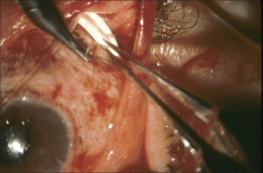
Eye undergoing glaucoma drainage tube surgery, from the Tube versus Trabeuclectomy study. (Source via CC by 2.0)
Surgical interventions for glaucoma are instituted when the patient does not respond to medical treatment. Glaucoma drainage implant (GDI) with a Baerveldt tube has been shown to have a higher success rate than trabeculectomy with mitomycin C, although the risk of corneal decompensation was found to be higher in the tube group. Other studies have also suggested that endothelial cell density (ECD) loss after GDI surgery is progressive.
As GDI surgery is becoming more popular in the treatment of glaucoma, this prospective clinical study was performed to determine the effect of GDI surgery on corneal topography parameters in the first postoperative year.
A total of 21 Caucasian patients were recruited in the study, excluding those with any form of corneal disease or previous related surgery (e.g. an anterior chamber intraocular lens, IOL). Previous uncomplicated surgery with an in-the-bag IOL, trabeculectomy, deep sclerotomy or vitrectomy was permitted in recruited patients. A total of 8 control eyes were found eligible. Patients received one of two kinds of implants (Molteno3, n=13, or Baerveldt 250, n=8). The final number of eyes included for analysis was at 20. Data was collected at different time points post-operatively until 12 months after.
Results showed that there was a statistically significant decrease in both the intraocular pressure (IOP) and in the number of anti-glaucoma medications that the patient was taking. The logMar visual acuity remained unchanged for most of the patients (14 eyes, 70%) and decreased in the others due to existing ocular conditions (e.g. progression of cataracts and macular degeneration).
At 1 year post-operatively, there was no difference in corneal astigmatism and keratometry between the eyes that have undergone GDI surgery and those that did not. There was an observed transient thickening of the central cornea at 6 months after surgery, which was not seen 12 months post-operatively. However, there was a significant decrease in ECD 1 year after surgery in both the central and peripheral cornea (8% and 9%, respectively). Patients with pseudoexfoliative glaucoma were observed to have a greater propensity to developing ECD changes after GDI surgery.
The researchers concluded that the findings of this pilot study has implications for IOL power calculations in patients who have to undergo cataract surgery after GDI. Further studies are necessary to evaluate the factors affecting GDI-surgery induced ECD loss.
Koivusalo, R., & Välimäki, J. (2019). Effect of glaucoma drainage implant surgery on corneal topography: a prospective study. Acta Ophthalmologica. doi: 10.1111/aos.14247