OCT imaging shows that it is significantly shorter in glaucomatous eyes versus healthy eyes.

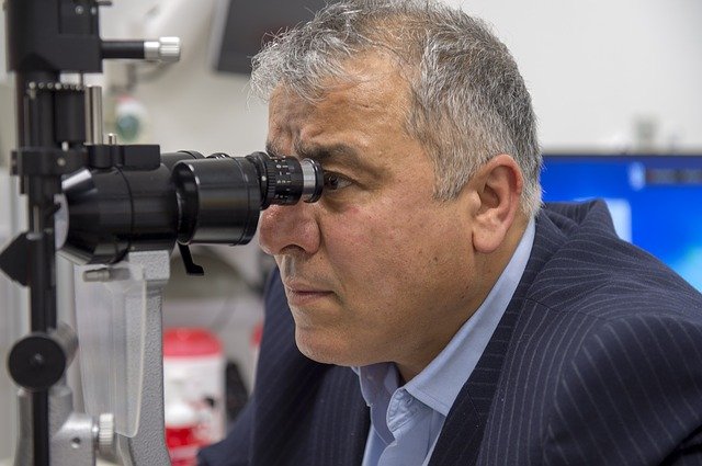
Intraocular pressure is best measured by applanation tonometry behind the slit-lamp.
Primary open angle glaucoma (POAG) is one of the leading causes of irreversible blindness worldwide. The clinical factor prognostic of progression is intraocular pressure (IOP), which is increased in glaucoma mostly due to outflow tract resistance near the Schlemm’s canal (SC).
A previous study in cadaver eyes has shown that in the eyes of patients with POAG, the scleral spur is shorter. It has been suggested that a shorter scleral spur would be less able to support an open SC. This study aims to compare the scleral spur length between diseased and healthy eyes using optical coherence tomography (OCT) imaging.
In this prospective clinical study, 78 patients 93 individuals with glaucomatous and healthy eyes, respectively, were recruited. The scleral spur was measured with three different methods, and the SC was imaged as well. These two parameters were observed in all four quadrants. Mean values were obtained for statistical analysis.
Results showed that using all methods, the length of the scleral spur was significantly shorter in POAG eyes than in healthy eyes. Used as a parameter, scleral spur length had good discriminating capability (AUC = 0.41). It was also found that the area of the Schlemm’s canal was significantly associated with the length of the scleral spur.
The study therefore concluded that scleral spur can be used as a diagnostic tool in differentiating eyes with POAG.
Li, M., Luo, Z., Yan, X., & Zhang, H. (2020). Diagnostic power of scleral spur length in primary open-angle glaucoma. Graefe’s Archive For Clinical And Experimental Ophthalmology. https://doi.org/10.1007/s00417-020-04637-4